Protection of Nervous Tissue
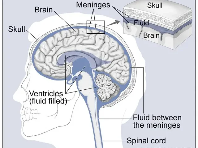
The Nervous Tissue in the brain and spinal cord is highly specialized and requires robust protection to ensure proper functioning and safety from injury. The protection is provided by several structures and mechanisms:
 >
>- Skull and Vertebral Column:
- Skull: The skull, a bony structure that serves as a rigid protective barrier against external mechanical forces, encases the brain.
- Vertebral Column: The spinal cord is housed within the vertebral column, which is made up of individual vertebrae. These bones encase the spinal cord, providing structural support and protection.
- Meninges:
- The brain and spinal cord are surrounded by three layers of protective membranes called meninges. These layers are:
- Dura Mater: The outermost, toughest layer that lies just beneath the skull and vertebrae.
- Arachnoid Mater: The middle layer, which has a web-like structure. It provides a cushioning effect.
- Pia Mater: The innermost layer that closely adheres to the surface of the brain and spinal cord, following their contours.
- The brain and spinal cord are surrounded by three layers of protective membranes called meninges. These layers are:
- Cerebrospinal Fluid (CSF):
- CSF is a clear fluid that circulates in the subarachnoid space (between the arachnoid mater and pia mater) and within the ventricles of the brain. It serves multiple protective functions:
- Cushioning absorbs shocks and protects the brain and spinal cord from mechanical injury.
- Buoyancy: This reduces the brain’s effective weight, preventing it from compressing under its own weight.
- Nutrient and Waste Transport: facilitates the transport of nutrients to nervous tissue and removes waste products.
- CSF is a clear fluid that circulates in the subarachnoid space (between the arachnoid mater and pia mater) and within the ventricles of the brain. It serves multiple protective functions:
- Blood-Brain Barrier (BBB):
- The BBB is a selective permeability barrier formed by endothelial cells of the brain capillaries. It prevents the passage of harmful substances from the bloodstream into the brain while allowing essential nutrients to pass through.
- Functions: Protects the brain from toxins, pathogens, and plasma composition fluctuations that could disrupt neural function.
How Nervous Tissue Causes Action
Nervous tissue, which includes neurons and glial cells, is critical to the nervous system’s function. It causes action by transferring electrical and chemical signals. Here’s how the process works:
 >
>- Neurons:
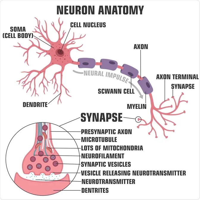
- Structure: Neurons consist of three main parts: the cell body (soma), dendrites, and axons.
- The cell body contains the nucleus and organelles, which are responsible for maintaining cell function.
- Dendrites are branched extensions that receive signals from other neurons.
- Axon: A long projection that transmits signals away from the cell body to other neurons, muscles, or glands.
- Structure: Neurons consist of three main parts: the cell body (soma), dendrites, and axons.
- Signal Transmission:
- Resting Potential: Neurons have a resting membrane potential, typically around -70 mV, maintained by ion pumps and channels.
- Action Potential: When a neuron is stimulated, ion channels open, causing a rapid influx of sodium ions (Na+), followed by an efflux of potassium ions (K+). This depolarization and subsequent repolarization create an electrical impulse called an action potential.
- Propagation: The action potential travels along the axon to the synaptic terminals.
- Myelination: Many axons are covered with myelin, a fatty substance produced by glial cells (oligodendrocytes in the CNS and Schwann cells in the PNS). Myelin sheaths speed up the transmission of action potentials through saltatory conduction, where the impulse jumps between Ranvier’s nodes (gaps in the myelin sheath).
- Synaptic Transmission:
- Synapse: The junction between two neurons or between a neuron and an effector cell (e.g., muscle cell).
- Neurotransmitter Release: When an action potential reaches the synaptic terminal, it triggers the release of neurotransmitters stored in vesicles into the synaptic cleft (the gap between the neurons).
- Receptor Binding: Neurotransmitters bind to receptors on the postsynaptic membrane, causing ion channels to open and generate a new action potential or response in the postsynaptic cell.
- Types of Actions:
- Voluntary Actions: Motor neurons initiate themselves in response to signals from the cerebral cortex. These actions involve skeletal muscles and are under conscious control.
- Involuntary Actions: Controlled by the autonomic nervous system (ANS) and involve smooth muscles, cardiac muscles, and glands. These actions are not under conscious control and include processes like heart rate, digestion, and respiratory rate.
Reflex Action
Reflex actions are rapid, involuntary responses to stimuli. They are controlled by the reflex arc and do not require conscious thought.
- Reflex Arc:
- Components:
- Receptor: Detects the stimulus (e.g., pain, heat).
- The sensory neuron transmits the signal from the receptor to the spinal cord or brainstem.
- Interneuron: processes the information and forms a response (located in the spinal cord or brainstem).
- The motor neuron transports the response signal from the spinal cord or brainstem to the effector.
- Effector: muscle or gland that performs the response action (e.g., moving a hand away from a hot object).
- Components:
- Types of Reflexes:
- Spinal Reflexes: Involves the spinal cord (e.g., knee-jerk reflex).
- Cranial reflexes involve the brain (e.g., blinking in response to a bright light).
Functions of Reflex Actions
- Protection: Reflexes protect the body from harm by enabling quick responses to potentially damaging stimuli (e.g., pulling a hand away from a hot surface).
- Homeostasis: Reflexes help maintain internal stability by regulating functions like heart rate, breathing, and digestion.
- Coordination: Reflexes assist in coordinating movement and posture.
Reflex Action through Spinal Cord and Brain
- Spinal Reflexes:
- Mechanism: Sensory neurons directly synapse with motor neurons in the spinal cord, enabling a quick response without involving the brain.
- Examples: Withdrawal reflex (pulling away from pain), patellar reflex (knee-jerk).
- Brain Reflexes:
- Mechanism: Sensory input is processed in the brainstem or higher brain centers before sending out a motor response.
- Examples: pupillary reflex (adjusting pupil size in response to light); gag reflex (preventing choking).
Understanding the protection and action mechanisms of nervous tissue allows us to appreciate how the nervous system efficiently maintains bodily functions and responds to the environment.

