Download the Cell organelles and their functions pdf is from the bottom of this article.
Cell organelles and their functions pdf
Protoplasm
Protoplasm is the essential living substance of a cell, encompassing all its components in a transparent, jelly-like form. Huxley defined it as the physical basis of life, while O. Hertwig’s protoplasmic theory (1892) emphasizes its role as the living matter in plants and animals. Divided into cytoplasm and nucleoplasm, protoplasm consists of key elements—carbon, hydrogen, nitrogen, and oxygen—along with both inorganic and organic substances.
Water makes up 90 percent of protoplasm, supplemented by mineral salts like sodium chloride and essential gases such as oxygen and carbon dioxide. Organic substances include proteins, carbohydrates, lipids, nucleic acids, and enzymes. Protoplasm performs vital functions such as nourishing, respiring, excreting waste, reproducing, metabolizing, and growing. It displays irritability and conductivity and is denser with higher viscosity than water. Its colloidal nature allows it to exist in both sol and gel states. The movement of protoplasmic molecules is known as Brownian movement, while the substances formed by it are termed “deutoplasm.” Understanding protoplasm deepens our appreciation of the fundamental processes that sustain life.

Functions of Protoplasm
- Metabolism: Site of biochemical reactions (catabolism and anabolism).
- Cellular Organization: Provides structural support and maintains cell shape.
- Transportation: Facilitates movement of molecules and organelles within the cell.
- Nutrient Storage: Stores essential nutrients (e.g., glycogen, lipids, proteins).
- Cell Division: Involved in mitosis and meiosis, including cytokinesis.
- Cell Communication: Contains signaling molecules for internal and external communication.
- Synthesis of Biomolecules: Involved in the synthesis of proteins and nucleic acids (RNA and DNA).
- Homeostasis: Regulates internal environment (pH, temperature, ionic concentrations).
- Defense Mechanisms: Supports immune responses in multicellular organisms.
- Energy Production: Site of cellular respiration, producing ATP for energy.
Cell wall
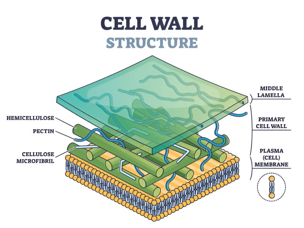
The cell wall is a rigid or semi-rigid structure located outside the cell membrane in plants, fungi, and most prokaryotic cells. It is this cell wall that creates a significant distinction between plant cells and other eukaryotic cells. The cell wall encases the protoplast, making it a part of the apoplast.
It provides structural support and protection while also functioning as a filtering mechanism. In fact, most of the carbon in terrestrial ecosystems is found in the cell walls of plants. In plants, the cell wall is primarily composed of cellulose, often combined with lignin. In fungi, the cell wall mainly consists of polysaccharides, while in bacteria, it is made up of peptidoglycan. Diatoms, on the other hand, have cell walls made of silicic acid.
Components of Cell wall
The cell wall consists of three main components: the middle lamella, the primary wall, and the secondary wall. In some cases, a tertiary cell wall may also be present.
- Middle Lamella: The middle lamella is the outermost layer that forms between adjacent cells during cell division. It is primarily composed of calcium and magnesium pectate compounds. This layer is viscous and serves as a cementing material, holding neighboring cells together.
- Primary Cell Wall: The primary cell wall is typically a thin, flexible, and extensible layer that forms while the cell is growing. The cellulose in the primary wall is stabilized by hydrogen bonds. Cellulose molecules create long chains that are interconnected with polysaccharide units known as pectin.
- Secondary Wall: As the cell matures, it develops an inner secondary wall layer, located just inside the primary wall. This secondary wall is approximately 5–10 micrometers thick and contains an additional compound called lignin, which makes the cell walls waterproof. The secondary wall is more rigid than the primary wall and provides additional support to the plant. It serves as the primary defense against harmful microbes, such as fungi and bacteria. The secondary wall is the main component of wood.
- In some instances, a tertiary wall may be found beneath the secondary wall, as seen in gymnosperm tracheids. This tertiary wall is fragile and primarily composed of hemicellulose xylan, lacking cellulose microfibrils.
Plasmodesmata
The term “plasmodesmata” was coined by Strasburger in 1901. The primary wall features tiny pores that allow the cytoplasm of adjacent cells to communicate through small channels known as plasmodesmata. Most plasmodesmata contain a narrow tube-like structure called a ‘desmotubule,’ which originates from the endoplasmic reticulum of the connected cells. Plasmodesmata are typically formed during cell division and are about 50–60 nanometers in diameter. A typical plant cell may contain between 103 and 105 plasmodesmata.
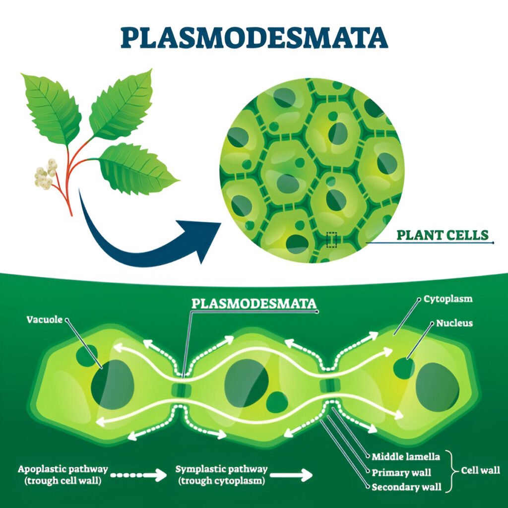
Plasmodesmata allow free movement of small metabolites as well as growth hormones between cells.
Symplast vs Apoplast
Due to the presence of plasmodesmata and intercellular spaces, the plant body is divided into two parts:
- Symplast: It is the living part of a plant body. It comprises a protoplast bounded by a single plasma membrane. It stores energy reserves.
- Apoplast: Apoplast is the nonliving part of a cell. It is external to the plasma membrane. It comprises a cell wall, spaces between cells,
Cell membrane
Every cell is surrounded by a thin, elastic membrane that isolates the cytoplasm from the external environment. This membrane is known as the cell membrane, which is also referred to as the plasma membrane or plasmalemma. The cell membrane is selectively permeable and cannot be seen under a light microscope. Its primary function is to regulate the controlled entry and exit of ions, such as sodium, potassium, and calcium.
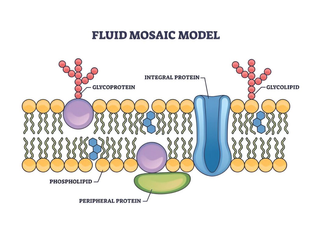
The cell membrane is present in both prokaryotic and eukaryotic cells. In bacteria, yeast, and plants, this membrane is bound by a cell wall. Typically, the thickness of the cell membrane is around 70–75 Å. Chemically, it is composed of lipids, proteins, and a small amount of carbohydrates. The arrangement of phospholipids, proteins, and carbohydrates in the plasma membrane is described by the fluid mosaic model.
Fluid mosaic model
According to the fluid mosaic model proposed by Singer and Nicolson in 1972, the cell membrane consists of a dynamic arrangement of lipids, proteins, and carbohydrates. The membrane features a continuous layer of phospholipids organized into a bilayer. The polar, hydrophilic phosphate heads face outward, while the nonpolar, hydrophobic fatty acid tails are oriented towards each other in the interior of the bilayer.
Proteins are embedded within this phospholipid bilayer, creating a mosaic structure. There are two types of proteins: extrinsic (peripheral) and intrinsic (integral). Extrinsic proteins can be easily removed, whereas intrinsic proteins are more firmly attached and not easily separable.
Due to the rapid movement of both protein and lipid molecules within the membrane, it is considered highly fluid. Additionally, the extracellular surface of the plasma membrane is adorned with carbohydrate groups (oligosaccharides), forming a loose coating known as the glycocalyx.
Role of Lipids
Lipid molecules are essential for maintaining the fluid properties of the cell membrane. The absence of covalent bonds between the lipids in the bilayer allows for fluidity in the membrane. Typically, the flip-flop movement of lipid molecules from one monolayer to the other occurs infrequently.
However, lipid molecules can quickly exchange places with their neighbors within the same monolayer, resulting in lateral diffusion. The presence of double bonds in unsaturated hydrocarbons tends to increase the fluidity of phospholipids.
In addition, inositol plays a significant role in cell signaling, while glycolipids assist in cell recognition. Furthermore, sterols help inhibit phase transitions in the membrane.
Role of Proteins
Proteins make up the primary structure of a cell membrane. Transport proteins play a crucial role in moving specific substances across the membrane. Channel proteins create open pores in the membrane, facilitating the passage of molecules. Carrier proteins selectively bind to and transport small molecules.
Many proteins also function as enzymes. Additionally, some peripheral proteins act as anchor points for the cytoskeleton or extracellular fibers.
Role of Carbohydrates
Oligosaccharides give a cell identity (i.e., distinguish self from non-self) and are a distinguishing factor in human blood types and transplant rejection.
They help in maintaining the asymmetry of a membrane.
It has been suggested that oligosaccharides are negatively charged, so the positively charged protein may remain bound to a cell membrane due to electrostatic attraction.
Intercellular Junctions
The plasma membrane undergoes modifications to create specialized structures that facilitate various functions, including absorption, fluid transport, electrical coupling, and cell adhesion. These modifications include:
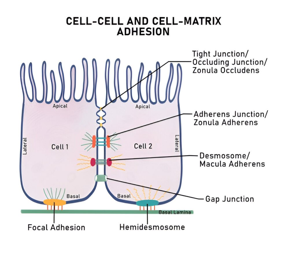
- Microvilli: Enhance absorption by increasing surface area.
- Tight junctions: Seal intercellular spaces to prevent leakage between cells.
- Desmosomes: Provide intercellular attachments that strengthen tissue integrity.
- Interdigitations: Increase adherence and surface area for the exchange of materials.
- Gap junctions: Enable intercellular communication by allowing molecules to pass directly between adjacent cells.
- Adherens Junctions: Composed of cadherin proteins linked to actin filaments, these junctions provide strong adhesion between adjacent cells, maintaining tissue integrity, especially in epithelial tissues.
- Focal Adhesions: Formed by integrins interacting with the extracellular matrix (ECM), focal adhesions anchor cells to the ECM and are crucial for signal transduction, cell migration, and response to mechanical forces, allowing dynamic interactions with the surroundings.
- Hemidesmosomes: These structures anchor epithelial cells to the basal lamina using integrins that connect to keratin filaments, providing stability and resistance to mechanical stress in tissues like skin.
These specialized structures play crucial roles in maintaining cellular functions and overall tissue organization.
Functions of Cell membrane
- Shape and Protection: The plasma membrane provides structural shape to the cell and protects the cytoplasmic organelles.
- Selective Permeability: It allows for the regulated movement of certain substances in and out of the cell.
- Transport Mechanisms: Molecules cross the membrane through active or passive transport processes.
- Hormone Recognition: The plasma membrane contains specific sites that facilitate the recognition of particular hormones.
- Membrane Fusion Regulation: It regulates the fusion of the membrane with other cellular membranes via specialized junctions.
- Enzyme Binding and Catalysis: The membrane provides specific sites for the binding and catalysis of enzymes.
- Secretion of Products: It aids in the release of secretory products from the cell.
Cytoplasm
- Location: The cytoplasm is the part of the cell located between the cell membrane and the nuclear membrane.
- Composition: It consists of cytosol (the matrix) and various organelles.
- Cytosol: The cytosol is a transparent, semifluid substance that exists outside of the organelles.
- Organelles: Organelles are membrane-bound structures within the cell, embedded in the cytoplasm.
- Characteristics of Organelles: Each organelle has a characteristic shape, specific chemical composition, and distinct functions.
- Types of Organelles: Organelles can be classified as:
- Extracytoplasmic: Such as the nucleus.
- Cytoplasmic: Including structures like mitochondria, plastids, Golgi complex, lysosomes, ribosomes, endoplasmic reticulum, centrosomes, cytoskeleton, cilia, and flagella.
- Function of Cytoplasm: The cytoplasm serves as the medium for various chemical reactions within the cell.
Plastids
- Plastids are large cell organelles found exclusively in autotrophic cells, such as those in plants and algae.
- Origin of the Term: The term “plastid” was introduced by botanist Andreas Schimper in 1885.
- Shape: Plastids are generally spherical or ovoid.
- Derivation: All plastids are derived from proplastids, which are immature plastids found in meristematic (actively dividing) tissues.
- Visibility: Plastids can be easily observed using a light microscope due to their size and distinct structures.
- Interconversion: Once formed, one type of plastid can be converted into other types based on the cell’s metabolic needs.
Types of Plastids

- Leucoplasts:
- Amyloplasts: Store starch and are primarily found in non-photosynthetic tissues, such as tubers and seeds.
- Elaioplasts: Store fats and oils, typically found in seeds and specialized cells.
- Aleuroplasts: Store proteins and are often found in seeds and storage tissues.
- Chromoplasts:
- They contain pigments (such as carotenoids) that provide color to fruits, flowers, and vegetables. They also attract pollinators and seed dispersers.
- Chloroplasts:
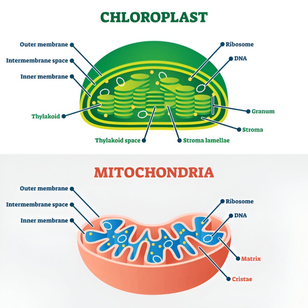
- Involved in photosynthesis, converting light energy into chemical energy (glucose) using chlorophyll. Chloroplasts are crucial for the plant’s ability to produce its food.
Functions of Plastids
- Energy Storage: Different types of plastids store various forms of energy, including starch (amyloplasts), fats (elaioplasts), and proteins (aleuroplasts) for later use by the plant.
- Photosynthesis: Chloroplasts are the primary site for photosynthesis, converting sunlight, carbon dioxide, and water into glucose and oxygen.
- Pigmentation: Chromoplasts contribute to the coloration of flowers and fruits, aiding in pollination and seed dispersal by attracting animals.
- Metabolic Pathways: Plastids are involved in various metabolic pathways, synthesizing and storing important metabolites such as fatty acids and amino acids.
- Developmental Role: Plastids play a role in plant development and differentiation, adapting their form and function to the plant’s needs.
Mitochondria
- Importance: Mitochondria are crucial cell organelles found in all eukaryotic cells, playing a vital role in energy production.
- Evolutionary Origin: They are descendants of free-living bacteria and account for about 20% of the cytoplasmic volume.
- Function: Mitochondria are the primary sites for cellular respiration and oxidative phosphorylation, where energy (ATP) is produced.
- Historical Discovery:
- First observed by Kolliker in 1850.
- Flemming (1882) referred to them as “fila.”
- Altmann (1892) named them “bioblasts.”
- Benda (1897) introduced the term “mitochondria,” derived from Greek words meaning “thread” and “granule.”
- Michaelis (1905) stained them using Janus Green B.
- Kingsbury (1912) identified them as the sites of cellular respiration.
- Palade and Sjöstrand (1940–1950) studied their fine structure.
- Size and Abundance:
- In animal cells, mitochondria are the second-largest organelle; in plant cells, they are the third largest.
- They are semi-autonomous organelles, containing their own DNA and machinery for protein synthesis.
- Mitochondria are the organelles that house the electron transport chain.
- Their lifespan is approximately 5–10 days.
- Absence in Certain Cells: Mitochondria are absent in prokaryotes and mature mammalian red blood cells (erythrocytes).
- Variability in Number: The number of mitochondria varies between individuals and among different cell types within the same individual, often correlating with metabolic activity. For example:
- Micromonas and Trypanosoma have one mitochondrion each.
- Sperm cells contain 20–24 mitochondria.
- Kidney cells have 300–400 mitochondria.
- Liver cells have 1,000–1,600 mitochondria.
- Insect flight muscles can contain approximately 500,000 mitochondria.
- Dynamic Numbers: The number of mitochondria can increase through fission (after mitosis) and decrease by fusion. Defects in these processes can lead to serious health issues.
- Shape and Size: Mitochondria have a variable shape, typically resembling a sausage. Their length ranges from approximately 1.5 to 10 micrometers, with a diameter of about 0.25 to 1 micrometer.
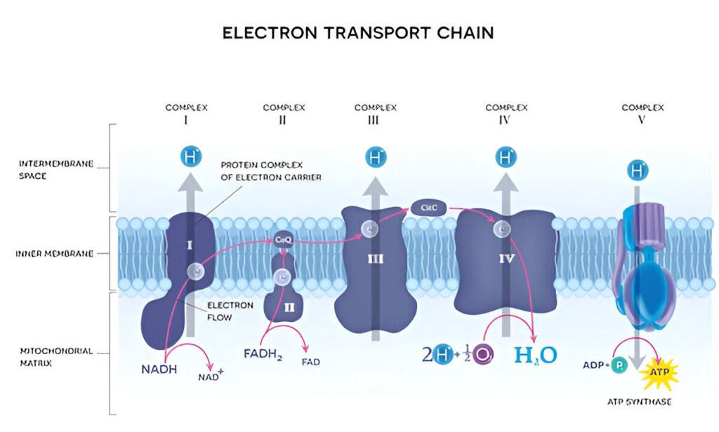
- Membrane Structure: Mitochondria are surrounded by a double membrane:
- Outer Membrane: Permeable to many metabolites and contains porin proteins and low levels of cardiolipin along with carrier proteins.
- Inner Membrane: Contains five complexes of integral membrane proteins involved in the electron transport chain:
- (a) NADH dehydrogenase (Complex I)
- (b) Succinate dehydrogenase (Complex II)
- (c) Cytochrome c reductase (Complex III)
- (d) Cytochrome c oxidase (Complex IV)
- (e) ATP synthase (Complex V)
- Intermembrane Space: The space between the outer and inner membranes is referred to as the outer chamber.
- The inner membrane contains stalked particles known as elementary particles or subunits of the Fernande–Moran complex (F0–F1 complex).
- Each elementary particle consists of three components: a head, a stalk, and a base.
- The head measures approximately 75–100 Å in diameter.
- The stalk is about 50 Å in length.
- The base ranges from 40 to 115 Å in size.
- The space between two elementary particles is roughly 100 Å, with an estimated 10^4 to 10^5 particles per mitochondrion.
- Chemical Composition:
- The head of the elementary particle is composed of ATPase.
- The stalk contains oligomycin sensitivity protein (OSCP).
- The base functions as a proton channel.
Mitochondrial DNA (mtDNA)
- Isolation: The first mitochondrial DNA was isolated from chickens in 1966.
- Copies: Mitochondria contain between 5 and 100 copies of DNA molecules.
- Structure: Mitochondrial DNA can be either circular or linear.
- Stability and Density: mtDNA is more stable and has a higher density compared to nuclear DNA.
- Base Composition: Mitochondrial DNA is rich in guanosine and cytosine bases.
- Human mtDNA: Human mitochondrial DNA comprises only 16,596 bases.
- Inheritance Pattern: Mitochondrial DNA does not adhere to the conventional rules of genetic inheritance; it is exclusively inherited from the mother.
- Mutations and Disease: Mutations in mitochondrial genes can lead to various diseases. Some known conditions associated with mtDNA include:
- Parkinson’s disease
- Huntington’s disease
- Leber’s optic neuropathy
- Kearns–Sayre syndrome
- Role in Ageing: Mitochondrial DNA is implicated in the ageing process.
Endoplasmic Reticulum
- The endoplasmic reticulum is a double-walled, zigzag structure that branches off from and is continuous with the outer membrane of the nucleus.
- Nomenclature: The term “endoplasmic reticulum” was introduced by Porter in 1953.
- Presence: Endoplasmic reticulum is found in all eukaryotic cells, with the exception of mature mammalian erythrocytes. It constitutes 30–60 percent of the total endomembrane system.
Types of Endoplasmic Reticulum
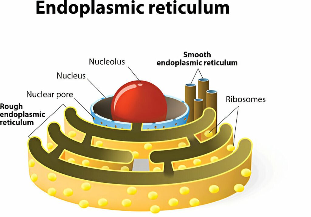
- Types of Endoplasmic Reticulum:
- Rough Endoplasmic Reticulum (RER): Studded with ribosomes, involved in protein synthesis and processing.
- STmooth Endoplasmic Reticulum (SER): Lacks ribosomes, involved in lipid synthesis, detoxification, and calcium storage.
- Specialized Forms:
- In muscle cells, the endoplasmic reticulum is referred to as the sarcoplasmic reticulum, primarily responsible for the storage and release of calcium ions (Ca²⁺).
- In retinal cells, it is known as myeloid bodies.
- Mobility and Elasticity: The endoplasmic reticulum is both mobile and elastic, allowing it to adapt to the needs of the cell.
- Structural Components:
- Cisternae: Flattened, tubular structures arranged parallel to each other, more abundant in rough endoplasmic reticulum, and interconnected.
- Vesicles: Oval to rounded structures that are abundant in secretory cells, involved in the transport of materials.
- Tubules: Unbranched or branched structures, abundant in cells that synthesize lipids.

Function of Endoplasmic Reticulum
- Mechanical Support: The endoplasmic reticulum provides mechanical support to the cell and contributes to the formation of the nuclear envelope.
- Detoxification: The smooth endoplasmic reticulum contains cytochrome P-450 enzymes, which detoxify pollutants, carcinogens, and drugs.
RER vs SER

Ribosomes
- Ribosomes are ribonucleoprotein particles and are among the smallest non-membranous cell organelles.
- Naming: They are named for their high RNA content and are commonly referred to as the “protein factory of the cell.”
- Historical Discovery: Ribosomes were first observed by Claude in 1941, who called them “microsomes,” while Palade coined the term “ribosomes” in 1958.
- Presence: Ribosomes are found in both prokaryotic and eukaryotic cells.
- Organelle Within Organelle: They are also known as organelles within organelles, as ribosomes are present in mitochondria and chloroplasts.
- Location: Ribosomes can exist in a free state in the cytoplasm or be attached to cytoplasmic membranes, such as the endoplasmic reticulum.
- Size: The average size of ribosomes is approximately 150–200 Å.
- Abundance: The number of ribosomes is particularly high in cells engaged in protein synthesis, such as pancreatic and liver cells.
- Functional Sites: Each ribosome has two functional sites:
- ‘A’ (Aminoacyl) Site: Receives tRNA.
- ‘P’ (Peptidyl) Site: Binds with the growing polypeptide-tRNA.
- Types of Ribosomes: There are two primary types of ribosomes:
- 70S Ribosomes: Found in prokaryotes and in mitochondria and chloroplasts.
- 80S Ribosomes: Found in eukaryotic cells.
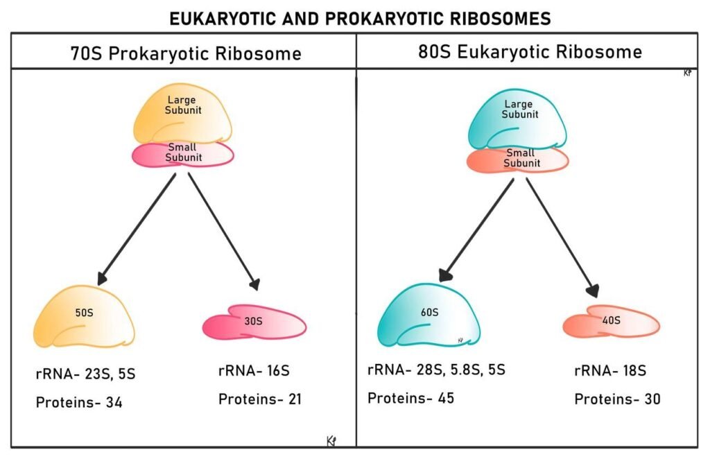
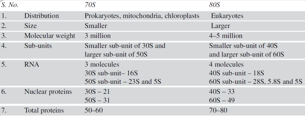
Difference between 70S and 80S Ribosomes
The main differences between 70S and 80S ribosomes are as follows:
- The association and dissociation of two sub-units of ribosomes depend on Mg++ ion concentration.
- In prokaryotes, biogenesis of ribosomes occurs in the cytoplasm, whereas in eukaryotes it occurs in the nucleolus.
- Ribosomes are the site of protein synthesis.
Lysosomes
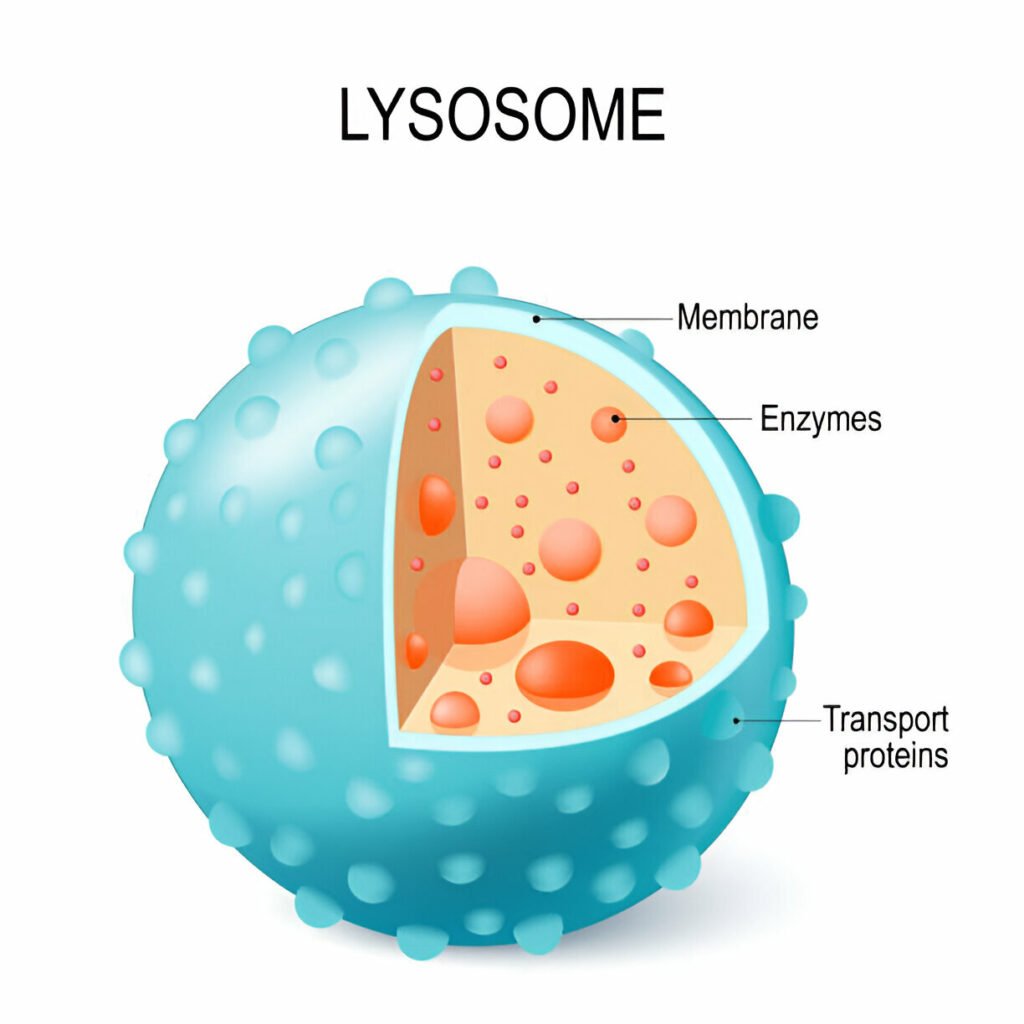
- Lysosomes are tiny organelles found in eukaryotic cells, often referred to as the “suicidal bags” of the cell due to their role in autolysis.
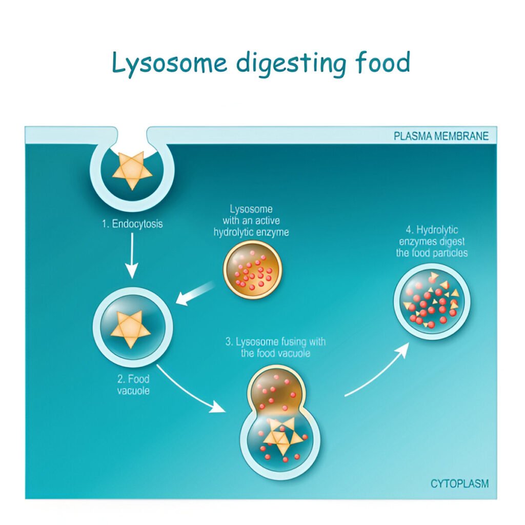
- Function: They are described as the “recycling center” of the cell because they digest worn-out organelles, freeing metabolites for reuse.
- Role in Cell Death: In many organisms, lysosomes are involved in programmed cell death.
- Nomenclature: The term “lysosome” was introduced by de Duve in 1955.
- Composition: Lysosomes are rich in hydrolytic enzymes and are surrounded by a single membrane.
- Digestive Capability: The enzymes within lysosomes can digest nucleic acids, polysaccharides, fats, and proteins, and most function optimally in an acidic environment.
- Stability: Lysosomes are stable within living cells, with their membranes resistant to the enzymes they contain.
- Membrane Stabilizers and Labilizers:
- Stabilizers: Cholesterol, cortisone, cortisol, and heparin help maintain lysosomal membrane integrity.
- Labilizers: Steroids, sex hormones, and lipid-soluble vitamins (A, D, E, and K) can weaken lysosomal membranes.
- Polymorphism: Lysosomes exhibit polymorphism, with four different types:
- Primary Lysosomes: Also known as storage granules, they contain digestive enzymes. Primary lysosome originates from the Golgi complex.
- Secondary Lysosomes: Also called heterophagosomes, formed by the fusion of primary lysosomes and engulfed materials.
- Autophagic Vacuoles: These lysosomes digest their own cell organelles.
- Residual Bodies: Contain undigested materials, representing the end stage of digestion.
- Disease Association: Malfunctioning lysosomes can lead to various diseases in humans, including:
- Tay–Sachs disease
- Niemann–Pick disease
- Farber’s disease
- Hunter’s syndrome
- Hurler syndrome
- Scheie syndrome
- Sanfilippo syndrome
- Digestion: Lysosomes are involved in both extracellular and intracellular digestion.
Golgi complex
- The Golgi complex is a cell organelle that exhibits structural variation and is primarily involved in cell secretion.
- Discovery: It was first identified by the Italian neurologist Camillo Golgi in 1898 while studying nerve cells in barn owls.
- Endomembrane System: The Golgi complex is a part of the endomembrane system and constitutes about 2 percent of the total cytoplasmic volume.
- Presence: It is found in all eukaryotic cells but is absent in prokaryotic cells, mature mammalian red blood cells (RBCs), and sperm cells.
- Variability: The shape and size of the Golgi complex can vary, depending on the physiological condition of the cells. It is particularly well-developed in secretory cells.
- Density: The specific density of the Golgi complex is lower than that of mitochondria and the endoplasmic reticulum.
- Ultrastructure Study: The ultrastructure of the Golgi complex was studied by Dalton and Felix in 1954 in the epididymis of rats.
Components of the Golgi Complex
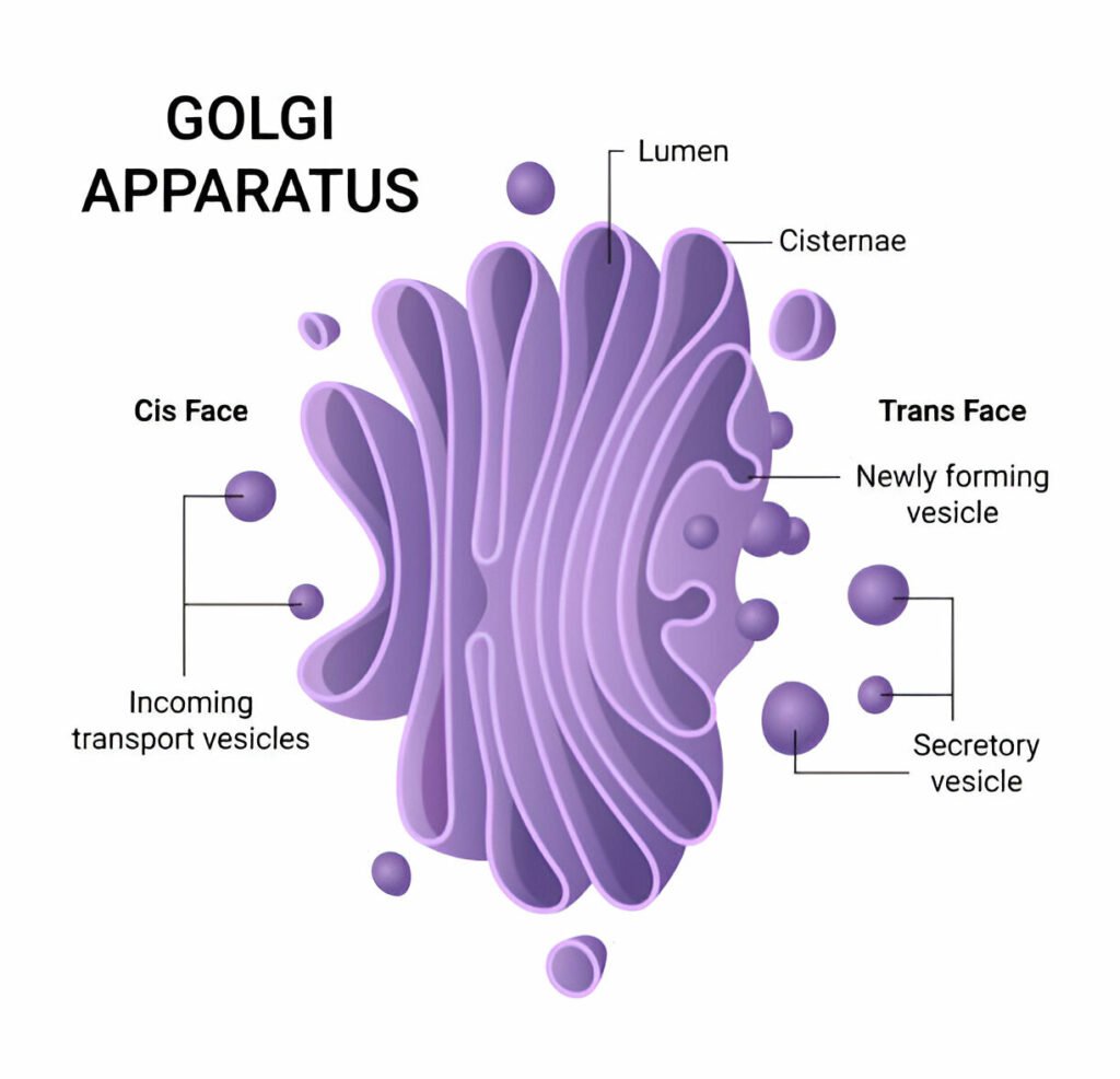
The Golgi complex is made up of three main components:
- Cisternae:
- Structure: Flattened, membrane-bound sacs that are stacked together.
- Function: Cisternae serve as the primary site for modifying, sorting, and packaging proteins and lipids received from the endoplasmic reticulum. Enzymes within the cisternae facilitate the glycosylation of proteins and the formation of lysosomal enzymes.
- Vesicles:
- Structure: Small, membrane-bound sacs that bud off from the cisternae.
- Function: Vesicles transport proteins and lipids to their appropriate destinations, either within the cell (e.g., to lysosomes) or outside the cell (e.g., through exocytosis). They play a crucial role in the secretion process.
- Vacuoles:
- Structure: Larger, membrane-bound compartments that can vary in size and content.
- Function: Vacuoles are involved in storage and transport within the cell. They can contain a variety of substances, including nutrients, waste products, and enzymes. In plant cells, vacuoles also help maintain turgor pressure.
Microbodies
- Microbodies are a diverse group of organelles found in the cytoplasm of almost all cells.
- Structure: They are roughly spherical structures bound by a single membrane.
- Functionality: Microbodies possess the ability to absorb oxygen and carry out direct oxidation.
- Nomenclature: The term “microbodies” was coined by Rohdin in 1954.
- Types: Microbodies can be mainly classified into two types: peroxisomes and glyoxysomes.
Peroxisomes
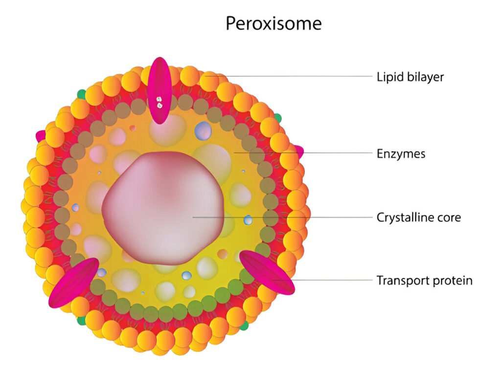
- Shape and Size: Peroxisomes are oval to round-shaped microbodies, found in all eukaryotic cells, with a diameter of approximately 0.5–1 µm.
- Replication: They are self-replicating organelles that bud off from the endoplasmic reticulum.
- Oxygen Utilization: Similar to mitochondria, peroxisomes are a major site of oxygen utilization.
- Enzymatic Content: They contain oxidative enzymes, such as catalase and urate oxidase, which facilitate various oxidative reactions.
- Function:
- Peroxisomes utilize molecular oxygen and hydrogen peroxide to perform oxidative reactions.
- In photosynthetic cells, they are involved in photorespiration.
- In animal cells, they play a crucial role in lipid metabolism.
- They protect cells from the toxic effects of hydrogen peroxide.
- Peroxisomes are particularly numerous in liver and kidney cells.
Glyoxysomes
- Glyoxysomes are a specialized form of peroxisomes found in certain plant cells, especially in germinating seeds and filamentous fungi.
- Shape and Size: They are oval, rounded, or polygonal microbodies, with a diameter of approximately 0.5–1 µm.
- Discovery: Glyoxysomes were first discovered and named by Briedenbach in 1967.
- Enzymatic Function: They contain enzymes responsible for the beta-oxidation of fatty acids and the glyoxylate cycle.
- Role in Metabolism: Glyoxysomes are involved in gluconeogenesis, converting stored lipids into carbohydrates during seed germination.

